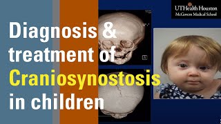
Craniosynostosis : Types, Causes, Diagnosis and Treatment : Pediatrics National exit test

Скачать Craniosynostosis : Types, Causes, Diagnosis and Treatment : Pediatrics National exit test бесплатно в качестве 4к (2к / 1080p)
У нас вы можете скачать бесплатно Craniosynostosis : Types, Causes, Diagnosis and Treatment : Pediatrics National exit test или посмотреть видео с ютуба в максимальном доступном качестве.
Для скачивания выберите вариант из формы ниже:
Cкачать музыку Craniosynostosis : Types, Causes, Diagnosis and Treatment : Pediatrics National exit test бесплатно в формате MP3:
Если иконки загрузки не отобразились, ПОЖАЛУЙСТА,
НАЖМИТЕ ЗДЕСЬ или обновите страницу
Если у вас возникли трудности с загрузкой, пожалуйста, свяжитесь с нами по контактам, указанным
в нижней части страницы.
Спасибо за использование сервиса video2dn.com


![EASY TRICK to Learn Congenital Heart Defects & Diseases [Pediatrics, Nursing, USMLE]](https://i.ytimg.com/vi/iT7-Flw_BTU/mqdefault.jpg)

Информация по комментариям в разработке