Candida on Sabouraud Dextrose Agar (SDA) typically forms colonies in yeast or pseudohyphal forms, with microscopic examination of saline wet mounts showing yeast-like cells and LPCB preparations revealing septate hyphae, true hyphae, and blastoconidia or pseudohyphae, depending on the specific Candida species and its ability to form these structures.
Candida on Sabouraud Dextrose Agar (SDA) typically forms colonies in yeast or pseudohyphal forms, with microscopic examination of saline wet mounts showing yeast-like cells and LPCB preparations revealing septate hyphae, true hyphae, and blastoconidia or pseudohyphae, depending on the specific Candida species and its ability to form these structures.
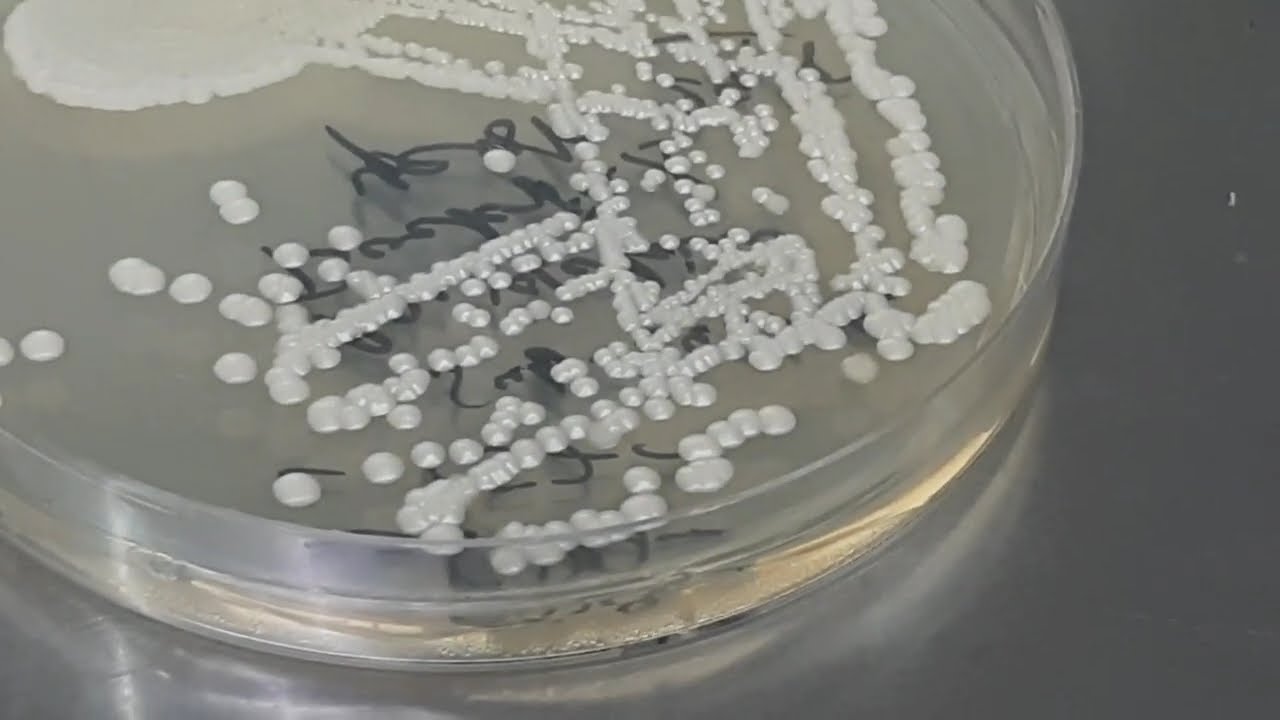
1. Candida Colony Morphology on SDA
Yeast form:
Colonies may be smooth, moist, and creamy, resembling bacterial colonies.
Filamentous (Hyphal/Pseudohyphal) form:
Some Candida species, such as Candida albicans, can also grow as filamentous structures, forming rough, matte colonies.
Color:
Colony color varies by species, but is generally white, cream, or yellowish.
2. Saline Wet Mount Microscopy
Purpose:
To observe the arrangement and morphology of fungal cells in a fresh or viable state.
Procedure:
A sample of the Candida culture is mixed with a drop of saline solution on a microscope slide and examined.
Microscopic findings:
This method will show yeast cells, which are typically oval or spherical. You may also observe pseudohyphae, which are elongated yeast cells that remain attached.
3. Lactophenol Cotton Blue (LPCB) Preparation
Purpose:
A staining method to visualize fungal structures and cell walls more clearly.
Procedure:
A small amount of the culture is placed on a slide with a drop of the LPCB solution, covered with a coverslip, and then examined under a microscope.
Microscopic findings:
Yeast cells: These appear as blue-stained, oval structures.
Pseudohyphae: Elongated, yeast-like cells in a chain, indicating a filamentous form.
Hyphae: True septate or non-septate hyphae, depending on the Candida species.
Blastoconidia: Conidia (spores) budding off from the yeast cells, or from the hyphal filaments.
Candida colony morphology, Candida species, Sabouraud dextrose agar, SDA growth, smooth creamy colonies, differential colony features, chromogenic media comparison, saline wet mount, budding yeast cells, pseudohyphae, germ tube negative, germ tube positive, chlamydospores, LPCB preparation, lactophenol cotton blue, microscopic demonstration, Candida albicans, Candida tropicalis, Candida glabrata, Candida parapsilosis, colony pigmentation, yeast identification, fungal microscopy, culture morphology, opportunistic fungi, clinical isolates, pathogenic yeasts, mycological diagnosis, fungal culture techniques, laboratory demonstration, microscopy findings

Информация по комментариям в разработке