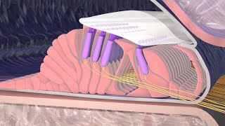
As sound waves enter the ear, they travel through the outer ear, the external auditory canal, and strike the eardrum causing it to vibrate. The central part of the eardrum is connected to a small bone of the middle ear called the malleus (hammer). As the malleus vibrates, it transmits the sound vibrations to the other two small bones or ossicles of the middle ear, the incus and stapes.
As the stapes moves, it pushes a structure called the oval window in and out. This action is passed onto the cochlea, which is a fluid-filled snail-like structure that contains the receptor organ for hearing.
The cochlea contains the spiral organ of Corti, which is the receptor organ for hearing. It consists of tiny hair cells that translate the fluid vibration of sounds from its surrounding ducts into electrical impulses that are carried to the brain by sensory nerves.
As the stapes rocks back and forth against the oval window, it transmits pressure waves of sound through the fluid of the cochlea, sending the organ of Corti in the cochlear duct into motion. The fibers near the cochlear apex resonate to lower frequency sound while fibers near the oval window respond to higher frequency sound.
Sound funnels into the ear canal and causes the eardrum to move. The eardrum vibrates with sound. Sound vibrations move through the ossicles to the cochlea. Sound vibrations cause the fluid in the cochlea to move. Fluid movement causes the hair cells to bend. Hair cells create neural signals which are picked up by the auditory nerve. Hair cells at one end of the cochlea send low pitch sound information and hair cells at the other end send high pitch sound information. The auditory nerve sends signals to the brain where they are interpreted as sounds. The outer ear collects sound waves moving through the air and directs them to the eardrum. The eardrum vibrates with sound. Sound vibrations move from the eardrum through the ossicles (bones in the middle ear) to the cochlea.
Hearing, or auditory perception, is the ability to perceive sound by detecting vibrations, changes in the pressure of the surrounding medium through time, through an organ such as the ear. Hearing mechanism. There are three main components of the human ear: the outer ear, the middle ear, and the inner ear.
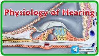
The outer ear includes the pinna, the visible part of the ear, as well as the ear canal which terminates at the eardrum, also called the tympanic membrane. The pinna serves to focus sound waves through the ear canal toward the eardrum. Because of the asymmetrical character of the outer ear of most mammals, sound is filtered differently on its way into the ear depending on what vertical location it is coming from. This gives these animals the ability to localize sound vertically. The eardrum is an airtight membrane, and when sound waves arrive there, they cause it to vibrate following the waveform of the sound. The middle ear consists of a small air-filled chamber that is located medial to the eardrum. Within this chamber are the three smallest bones in the body, known collectively as the ossicles which include the malleus, incus and stapes (sometimes referred to colloquially as the hammer, anvil and stirrup respectively). They aid in the transmission of the vibrations from the eardrum to the inner ear.
The purpose of the middle ear ossicles is to overcome the impedance mismatch between air and water, by providing impedance matching. Also located in the middle ear are the stapedius and tensor tympani muscles which protect the hearing mechanism through a stiffening reflex. The stapes transmits sound waves to the inner ear through the oval window, a flexible membrane separating the air-filled middle ear from the fluid-filled inner ear. The round window, another flexible membrane, allows for the smooth displacement of the inner ear fluid caused by the entering sound waves.
The inner ear consists of the cochlea, which is a spiral-shaped, fluid-filled tube. It is divided lengthwise by the organ of Corti, which is the main organ of mechanical to neural transduction. Inside the organ of Corti is the basilar membrane, a structure that vibrates when waves from the middle ear propagate through the cochlear fluid – endolymph. The basilar membrane is tonotopic, so that each frequency has a characteristic place of resonance along it. Characteristic frequencies are high at the basal entrance to the cochlea, and low at the apex. Basilar membrane motion causes depolarization of the hair cells, specialized auditory receptors located within the organ of Corti.
While the hair cells do not produce action potentials themselves, they release neurotransmitter at synapses with the fibers of the auditory nerve, which does produce action potentials. In this way, the patterns of oscillations on the basilar membrane are converted to spatiotemporal patterns of firings which transmit information about the sound to the brainstem.


Информация по комментариям в разработке