The first in a series using MRI next to my 3D models, aiming to simplify the 3D comprehension of the anatomy of the brain.
View this model here: https://www.aboutmedicine.com.au/mode...
MRI citation: Puccio et al.
--
In this video, we’re going to compare MRI images of the brain to this schematic 3D model.
By doing so, we’ll get a better intuition for how the parts of the brain fit together.
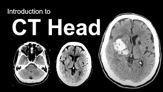
The three sets of images you see here are taken in the coronal, sagittal and transverse planes.
This corresponds to these planes on our 3D model - coronal, sagittal and transverse.
Throughout the video, I’ll be moving through all three planes.
Notice how, if I click and drag on this image, we move through the slices on the other planes.
Take a look at the coronal plane, as I drag the cursor through the sagittal plane, from right to left.
We’ll bring that back to the centre. Now look at the transverse plane images, as I drag the cursor from the top of the brain downward. This is also known as the axial plane.
Occasionally I’ll enlarge one plane like so. This is a sagittal plane image, in the midline, so we are looking at the very middle of the brain here.
Now let’s look at our 3D model for a moment.
To put it in context, we have this 2D body outline, but let’s remove that now.
To really ground ourselves, let’s recreate the image on the left. So we’ll take away half of all the models. And then we can line up our sagittal image.
Ok, now that you have a sense of perspective, let’s begin talking about the anatomy.
The first thing we see is the cortex - the grey matter on the outside of the brain.
Looking over to our MRI now, see the darker grey cortex here. It’s all bumpy and knobbly. Which is why it goes by the name cortex, meaning bark in Latin, because anatomists from back in the day thought it felt like the bark of a tree.
The cortex is divided into lobes, which we’ll just go through briefly, that’s the frontal, parietal, occipital and temporal lobes.
Beneath the grey matter, moving over to look at our MRI again, is all this white matter.
White matter is the axons, or the electrical wires, of the neurons that make up the brain.
The cell body, the part of the neuron that generates or manipulates information, is in the cortex.
Now, in order to move our muscles a message has to be generated in the brain and sent down the spinal cord.
Let’s think about the passage of that information in the brain.
So the cell body in the motor cortex, which sits in the back part of the frontal lobe, generates the message.
It sends it down the axon, part of the white matter here. The axon passes through this area, called the internal capsule.


Информация по комментариям в разработке