This video is about the Connective Tissue
Content:
Introduction 0:00
Development of CT 0:50
Classification of Connective Tissue 3:51
Extracellular Matrix 6:10
Fibroblasts 8:00
Dense Connective Tissue 8:50
Dense Regular Connective Tissue 9:30
What are Collagen Fibers made up of? 11:11
Suffixed of Cells -Blast, -Cyte, -Clast 12:06
Table for Collagen Fibers Types 13:25
Elastic Fibers 16:43
Reticular Fibers 17:40
Ground Substance 20:22
Cells in Connective Tissue 21:59
Macrophage 22:15
Mast Cell 23:20
Plasma Cells 24:22
White Adipocytes 25:30
Brown Adipocyte 26:48
Quiz Yourself 27:31
Follow my new Instagram: @Taimtalksmed
☕Support me at:
https://www.buymeacoffee.com/meditay
Development of Connective Tissue (Mesenchyme)
Zygote - Blastula - Gastrula
Gastrula becomes:
Ectoderm- Skin and nervous system
Endoderm
- GI tract, Glands, Respiratory tract
Mesoderm
- Mesenchyme
- Mesothelium (Membrane of Simple squamous epithelium becomes different body cavities)
Mesenchyme is connective tissue, connective tissue derives from the middle germ layer, mesoderm.
Mesenchyme:
- Bone, Cartilage, Tendons, Capsules, Ligaments, Hematopoietic cells
- Mesenchymal cells (Nuclei, Cell processes, connect and form a network called Syncytium)
- Mesenchyme is an embryonic tissue
- You find the mesenchyme under a microscope near the neural tube during development.
Classification of Connective Tissue
Common origin (Mesenchyme)
- CT Proper (Loose CT, Dense CT)
- CT with special properties (Adipose Tissue, Hematopoietic cells (Fluid CT), Elastin, Mucous issue)
- Supportive CT (Cartilage, Bone)
- Composition of connective tissue under basement membrane (Cells and extracellular matrix)
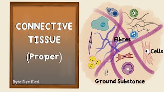
Extracellular Matrix
Consist of:
- Fibers (Collagen fibers, Elastin fibers, Reticular fibers)
- Ground Substance
- Extracellular Fluid
Fibroblasts and how to find them:
- Fibroblast and Reticulocytes
- Fibrocytes synthesize proteins
- Myofibroblasts, wound healing
- Fibroblasts have many forms, different cell processes
Dense CT Regular Dense Connective tissue (Tendons)
Irregular dense connective tissue (deep layer of dermis)
Dense regular connective tissue in tendons:
Consist of dense and loose connective tissue
Dense: Collagen fibers pressed together, fibroblast pressed together, elongated nuclei, called tendobast
Loose: Loose connective tissue with round fibroblast
Longitudinal and crossed histology section slide of Tendons have up to 10 orders:
1st order: between two tendoblasts
2nd order: between two loose connective tissue across dense connective tissue
Collagen fibers composition:
Collagen fiber, Fibril, Microfibril, Tropocollagen Helix
Suffixes in connective tissue cells
- Blast (Fibroblast, Osteoblast, Chondroblast)
- Cyte (Fibrocyte, Osteocyte, Chondrocyte)
- Clast (Fibroclast, Osteoclast, Chondroclast)
Collagen fibers types:
Type 1 collagen: (resistant to tention)
Type 2 collagen: (resistant to pressure)
Type 3 collagen: (maintenance in organs)
Type 4 collagen: (supports basal lamina)
Type 5 collagen: (resistant to tension, together with type 1)
Type 7 collagen: (connects basal lamina with reticular lamina, together with type 4 collagen)
Type 9 collagen: (together with type 2)
Type 10 collagen: (support bones, by chondrocytes)
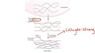
Elastic Fibers:
- Oxytalan: consist of strong fibrillin twines
- Elaunin fibers: elastin and fibrillin
- Proper elastic fibers: elastin and fibrillin
Reticular fibers:
Consist of Type 3 collagen
From reticulocyte
how to find the reticular fibers in a microscope:
Histology slide of lymph node
Zoom in to light area, reticulocyte with lymphocyte
Reticulocyte (Light nuclei, cell processes, bigger amount of cytoplasm than lymphocyte)
Challenge, try to find reticulocyte
Ground Substance
- between cells and fibers
- Acts as lubricants for cells and fibers and barrier for invaders.
- Glycosaminoglycans
- Proteoglycans
- Multi-adhesive glycoprotein
- Matrix metalloproteinase (type of proteinase)
How to find ells in connective tissue histology
Macrophage (from bone marrow, circulates as monocyte, becomes macrophage in tissue. bean-shaped nucleus)
Mast cell (large cell, has granules containing histamine and heparin) in microscope
Plasma cell (arise from B-Lymphocytes) Cartwheel nucleus, how to find it in histology slide. Light center of nucleus and dark in edges
White Adipocyte: Lipid droplet in cytoplasm. Energy storage (Lipid storage). Nucleus at side, cytoplasm at sides. Adipocyte subcutaneous tissue. Primary fat we have in the body.
Brown Adipocyte: Fat we have when we are born, no brown fat as adults. They look brown in a microscope because they have more mitochondria in the cytoplasm.
Challenge at the end of the video!


![Connective Tissue Fibers Histology [Connective Tissue Histology Part 1 of 3]](https://i.ytimg.com/vi/R9XCsQSoATE/mqdefault.jpg)


Информация по комментариям в разработке