The development of the pharyngeal arches (head and neck) explained in a very simple way.
If you are completely new to embryology and you want to understand it quickly, this should be the first video you watch:
• Introduction to Embryology - Fertilis...
Post any questions you have about the video below, I read all the comments:
--------------------------------
Recommended Text
--------------------------------
Easy Embryology is a book that is dedicated to the simplification of embryology. It is available at https://drminass.com/product/easyembr.... Contact Dr. Minass for more information.
----------------------------------------
Interact With Dr. Minass!
----------------------------------------
Website - https://www.drminass.com
Email - [email protected]
Patreon - / drminass
Facebook - / m1na55
Instagram - @m1.nass
Post - Address to:
Minass
Parcel Locker 10106 04448
59 Penshurst Street
Willoughby, NSW
Australia 2068
Summary for your notes:
HEAD AND NECK
The formation of the head region is derived from paraxial and lateral plate mesoderm, neural crest and thickened regions of ectoderm known as ectodermal placodes.
The most distinctive feature in the development of the head and neck is the presence of pharyngeal arches.
The pharyngeal arches appear in the fourth and fifth weeks of development and contribute to the characteristic external appearance of the embryo.
They initially consist of bars of mesenchymal tissue separated by deep clefts known as pharyngeal clefts.
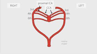
With the development of the pharyngeal arches, a number of outpocketings, the pharyngeal pouches appear along the lateral walls of the pharynx, the most cranial part of the foregut (for future video).
The pouches penetrate the surrounding mesenchyme but do not establish an open communication with the external clefts
Pharyngeal arches contribute to the formation of the neck and the face.
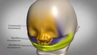
At the end of the forth week the centre of the face is formed by the stomodeum, surrounded by the first pair of arches.
At 42 days, five mesenchymal prominences can be recognised: mandibular prominence (first pharyngeal arch and caudal to stomodeum), maxillary prominences (dorsal portion of the first pharyngeal arch), frontonasal prominence (cranial to stomodeum). Development of the face is later completed by formation of the nasal prominences.
Pharyngeal arches
Each arch consists of a core of mensenchymal tissue covered on the outside by surface ectoderm and on the inside by epithelium of endodermal origin.
In addition to mesenchyme derived from paraxial and lateral plate mesoderm, the core of each arch receives substantial numbers of neural crest cells which migrate into the arches to contribute to skeletal components of the face.
The original mesoderm of the arches gives rise to the musculature of the face and neck. And these muscular components of each arch have their own cranial nerve and wherever muscle cells migrate, they carry their nerve component with them.
Additionally, each arch has its own arterial component, cranial nerve and cartilage. Listed below for your convenience :)
------------------------------------------------------------------------
WHAT THE PHARYNGEAL ARCHES BECOME
------------------------------------------------------------------------
ARCH 1: Maxillary and mandibular processes
Trigeminal nerve: maxillary and mandibular branches
Muscles: Mastication (temporal; masseter, medial, lateral pterygoids); mylohyoid, anterior belly of digastric, tensor palatine, tensor tympani
Premaxilla, maxilla, zygomatic bone, part of temporal bone, Meckel’s cartilage, mandible malleus, incus, anterior ligament of malleus, sphenomandibular ligament
ARCH 2: Hyoid
Facial nerve
Muscles of facial expression (buccinator, auricularis, frontalis, platysma, orbicularis oris, orbicu- laris oculi) posterior belly of digastric, stylohyoid, stapedius
Stapes, styloid process, stylohyoid ligament, lesser horn and upper portion of body of hyoid bone
ARCH 3:
Glossopharyngeal nerve
Stylopharyngeus
Greater horn and lower portion of body of hyoid bone
ARCHES 4–6
Vagus nerve
Superior laryngeal branch (nerve to fourth arch)
Recurrent laryngeal branch (nerve to sixth arch)
Cricothyroid, levator palatine, constrictors of pharynx
Intrinsic muscles of larynx
Laryngeal cartilages (thyroid, cricoid, arytenoid, corniculate, cuneiform)


Информация по комментариям в разработке