(USMLE topics, cardiology) Causes, symptoms and pathology of PAC.
Purchase a license to download a non-watermarked version of this video on AlilaMedicalMedia(dot)com
Check out our new Alila Academy - AlilaAcademy(dot)com - complete video courses with quizzes, PDFs, and downloadable images.
©Alila Medical Media. All rights reserved.
Voice by Ashley Fleming
All images/videos by Alila Medical Media are for information purposes ONLY and are NOT intended to replace professional medical advice, diagnosis or treatment. Always seek the advice of a qualified healthcare provider with any questions you may have regarding a medical condition.
Premature atrial contractions, PACs, are premature heartbeats originating in one of the upper chambers of the heart, the atria. PACs are common among patients with lung disorders, such as chronic obstructive pulmonary disease, COPD, but they also often occur in healthy people. PACs may be caused or worsened by caffeine, alcohol use, and certain medications. Apart from occasional palpitations, PACs are generally asymptomatic and do not require treatment in otherwise healthy people.
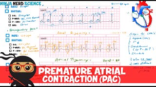
A PAC happens when the atria are activated by an ectopic site in an atrium, instead of the SA node. Because atrial depolarization is initiated outside the SA node, the associated P wave has an unusual, non-sinus morphology. An early atrial activation may cause the P wave to merge with the preceding T wave producing a peaked fusion wave. In situations where the ectopic site is located near the AV node, the atria are depolarized mainly by retrograde conduction and the resulting P wave is inverted; the PR interval representing the time the signal reaches the AV node, is slightly shorter. An ectopic atrial activation can usually enter the SA node, depolarize it and reset its timing, causing a so-called non-compensatory pause. On an ECG, this is seen as changes in the PP intervals that contain the ectopic beats. This feature can be used to differentiate PACs from ectopic beats of ventricular origin, PVCs. PVCs typically do NOT conduct back to the atria, SA node firing is NOT affected, and PP interval remains unchanged.
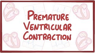
The downstream ventricular conduction of a PAC can be normal, aberrant or absent. In most cases, conduction through the AV node and ventricles is not affected, resulting in a normal narrow QRS complex. Occasionally however, ventricular conduction may be aberrant, causing a widened QRS complex, usually with right bundle branch block morphology, which may look similar to a premature beat of ventricular origin. In this case, differentiation is made based on the presence of a preceding P wave and a non-compensatory pause in PAC.
A non-conducted PAC is one that arrives too early to the AV node, at the time when the AV node is still in refractory period and thus cannot be activated.


Информация по комментариям в разработке