https://kunskapsprovet.com
Instagram: / kunskapsprovet
Facebook: / kunskapsprov
Telegram: https://t.me/joinchat/Bi7pEhjrR6MNmVb...
Whatsapp: https://chat.whatsapp.com/C1QkYjSls9x...
Youtube: / @kunskapsprov5884
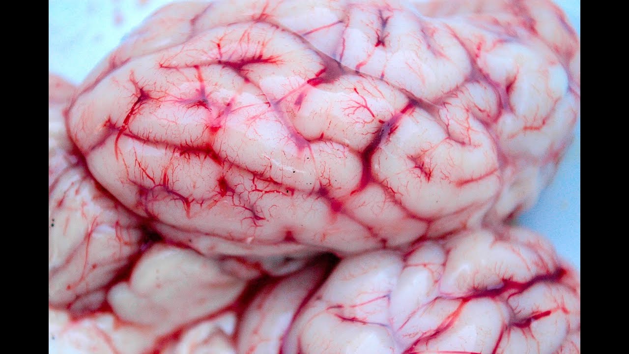
The human brain has a highly complex structure. It contains billions of neurons wired together through trillions of connections. Each portion of the brain has a distinct set of functions. Damage to a part of brain results in characteristic clinical manifestations. Knowledge of neuroanatomy, functions of different parts of the brain and set of clinical manifestations resulting from damage to a part of the brain is of paramount importance in the localization of a neurological lesion. Medical students are often perplexed with the complexity of this knowledge. This video lecture on the localization of a neurological lesion is aimed at delivering a succinct and easy to understand overview of the subject.[1][2][3][4]
Function
The nervous system is divided into the central nervous system (CNS) and peripheral nervous system. The central nervous system comprises of the brain and spinal cord. Cranial and spinal nerves make up the peripheral nervous system.
The brain consists of 2 cerebral hemispheres, brain stem, and cerebellum. The cerebral cortex is a convoluted structure that has multiple tortuous folds called gyri separated by deep grooves called sulci. Central sulcus separates frontal lobe from the parietal lobe, and the Sylvian fissure marks the upper boundary of the temporal lobe. An arbitrary line separates occipital lobe from parietal and temporal lobes.
Precentral gyrus serves as primary motor cortex and is the command and control center for voluntary movements. Pyramidal cells in the layer V of cerebral cortex innervate lower motor neurons located in the cranial and spinal motor nuclei through corticobulbar and corticospinal tracts, respectively. These pyramidal cells are called upper motor neurons. The distribution of these pyramidal cells follows a unique topographic pattern. Upper motor neurons that control lower motor neurons of the lower limb are located on the medial side, and those innervating lower motor neurons of the upper limb are located laterally. An area on the lateral surface of the dominant frontal lobe, the left frontal lobe in most individuals, is the motor control center for speech. It is called Broca’s area. Another area at the junction of parietal and temporal lobes analyzes sensory input related to speech and is called Wernicke's area. Brain stem consists of the midbrain, pons, and medulla. Some cranial nerves leave brain stem.
Descending fibers of corticospinal tract travel from the cerebral cortex to corona radiata, posterior limb of the internal capsule, cerebral peduncles, pons, and medulla. At the lower part of the medulla, most of these fibers cross the midline, continue as the lateral corticospinal tract and descend through the white matter of the cord to innervate the anterior horn cells. This crossing over of corticospinal tract fibers is called pyramidal decussation. Resultantly, pyramidal cells of right cerebral cortex innervate left spinal motor nuclei and vice versa. Spinal motor nuclei innervating skeletal muscles of upper and lower limbs receive upper motor neuron innervation from the contralateral side only. Lower motor neurons of cranial nerves, on the other hand, are innervated by corticobulbar fibers from both sides. Therefore despite damage to corticobulbar fibers on one side, cranial motor nuclei will continue to receive upper motor supply from the other side. The nucleus of the facial nerve, however, can be considered a hybrid. The upper half of facial nucleus, like other cranial motor nuclei, receives bilateral upper motor neuron innervation. On the other hand, the lower half receives innervation only from the contralateral side.
Medulla oblongata passes through the foramen magnum and continues as the spinal cord. The spinal cord has multiple segments. Each segment gives away a pair of spinal nerves. There are 8 cervical, 12 thoracic, 5 lumbar, 5 sacral, and 2 coccygeal segments. Looking at the cross-section, the spinal cord has a central gray matter that contains cell bodies of spinal nuclei and peripheral white matter that contains myelinated axons. Anterior horn of the gray matter contains spinal motor nuclei; axons of these neurons innervate skeletal muscles and are called lower motor neurons. Axons of these neurons travel through the anterior root of the spinal nerves and form brachial and lumbosacral plexuses that innervate upper and lower limbs, respectively.
Localization of Lesions; complete lecture. Contributed by Muhammad Zaman Khan, MD
https://www.ncbi.nlm.nih.gov/books/NB...

Информация по комментариям в разработке