Learning the Anti-Arrhythmic Agents just got a whole lot easier!
**MedImmersion to the rescue**
Listen guys, I really hope this video helps you in school. I definitely had fun making it! If you have questions, comments, or even criticisms...please, leave a comment. I love teaching and working with students, so your comments mean a lot to me!
Good Luck in school!
Hey YouTube, this is Dr. Joel.
In this video, I'm gonna be covering the antiarrhythmic agents.
I'm gonna start with a review of cardiac physiology, and then jump right into the agents themselves.
I'll cover the Class I, Class II, Class III, Class IV, Class V, and then just give you some departing thoughts and then I will finish off with a couple of knowledge challenge questions, just to see where you're at.
OK? Let's get started.
In order to do a really good review of the cardiac antiarrhythmic agents, it's first important important for me to cover a little bit of cardiac physiology, starting first with the cardiac action potential.
And that's because this action potential is a little bit different than the action potential that you're going to see in nerves.
Also, a solid understanding of this action potential will help you later understand why the drugs work the way they do.
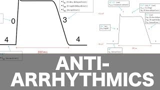
So, this picture on the right represents a cardiac action potential.
And, one thing that you need to understand is that this action potential is going to be a little bit different depending on which part of the heart you're measuring.
However, the principles that I'm about to cover will apply to all of those tissues in the heart.
And, if you want to, you can click on this link, which will take you to a picture that I think does a really cool job about showing the differences in the cardiac action potential in the different sections of the heart and that also how all those electrical depolarizations add up to make the electrocardiogram wave form.
Anyway, on the X axis, we have time and on the Y axis, we have voltage.
In the polarized state, the heart rests at about negative 95 millivolts.
An action potential cycle takes about 200 milliseconds.
And that number changes depending on which part of the heart you're in or which tissue you're sampling.
So, on this graph, you can see that the heart starts at about negative 95 millivolts then it very quickly shoots up to about 20 or so, by this graph, pause at 20 millivolts.
It stays there for a bit, and then the cell starts to repolarize itself.
And that's the cycle.
I'm going to add a cell membrane at the top of this picture and I'm going to walk through the phases of the action potential one at a time and what I want you to do is, I want you to imagine that above this cell membrane is the extracellular space and below this membrane is the intracellular space.
OK, starting off with Phase 0, which is the depolarization phase.
This is caused by a opening of voltage-gated sodium channels.
And these are very fast, rapid-acting channels that allow a large amount of sodium to move very quickly.
Sodium is positively charged, so if positive things come into the cell, then the cell becomes more positive.
OK, does that make sense? Basically, that's why you see this huge skyrocketing here of the voltage from negative 95 to positive 20.
It's because those positive sodium ions are moving in very quickly.
Next is Phase 1, which is the initial repolarization phase, which is basically caused by the rapid inactivation of those sodium channels.
Almost as quickly as they open, they start to close again.
At the same time, voltage-gated potassium channels start to open allowing potassium to efflux or exit the cell.
Potassium is also positively charged.
So if you have positive things leaving the cell, then the cell becomes more negative, right? And that's why there's a little dip there in the voltage.
Next, with Phase 2, you get calcium channels and they begin to open.
Calcium, again, also positive.
Positive things coming into the cell would make the cell more positive.
But potassium is still moving out, so that would make the cell more negative, and hence you get this plateau phase.
It kind of balances out for a little bit.
It's not exactly flat, but it's close.
We still call it the plateau phase.
And, as you know, the calcium plays an effect on how the muscle cells contract.
So that's important as well for contraction.
Next is the rapid repolarization phase, which is Phase 3.
More of the voltage-gated slow potassium channels are opening and they allow more potassium to rush out and the calcium channels begin to close so the cell starts to move back down to a negative value, a strong negative value.
And you have to remember, the sodium-potassium ATPase pump is also chugging along this whole time.
It's still working, it's still pumping potassium in and sodium out, which is just another factor that is driving that cell back down to its polarized state.
Lastly is the fourth phase, which is the resting potential phase.


Информация по комментариям в разработке