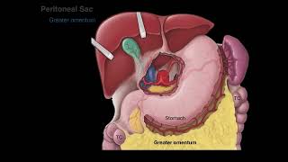
Tracing the peritoneum in sections - simplified

Скачать Tracing the peritoneum in sections - simplified бесплатно в качестве 4к (2к / 1080p)
У нас вы можете скачать бесплатно Tracing the peritoneum in sections - simplified или посмотреть видео с ютуба в максимальном доступном качестве.
Для скачивания выберите вариант из формы ниже:
Cкачать музыку Tracing the peritoneum in sections - simplified бесплатно в формате MP3:
Если иконки загрузки не отобразились, ПОЖАЛУЙСТА,
НАЖМИТЕ ЗДЕСЬ или обновите страницу
Если у вас возникли трудности с загрузкой, пожалуйста, свяжитесь с нами по контактам, указанным
в нижней части страницы.
Спасибо за использование сервиса video2dn.com


![Anatomical Position and Directional Terms [Anatomy MADE EASY]](https://i.ytimg.com/vi/t6-ueqFK1IE/mqdefault.jpg)

![Body Cavities and Membranes: Drawn and Defined [Anatomy Physiology]](https://i.ytimg.com/vi/sqEDTVYq1_U/mqdefault.jpg)
Информация по комментариям в разработке