Jennifer Alston DeSimone, MD, FAAD, Associate Professor, University of Virginia School of Medicine; Assistant Professor, Georgetown University Hospital; Director, Cutaneous Lymphoma and High Risk/Transplant Dermatology; INOVA Schar Cancer Institute Melanoma and Skin Oncology Center discusses the criteria to diagnose mycosis fungoides.
The diagnosis of CTCL is often challenging; as a result, delays in diagnosis (and subsequently work-up and treatment) can be significant. Part of the reason is the variability in how individual patients present with CTCL and its subtypes. Because mycosis fungoides progresses slowly, some patients may not experience progression beyond their initial symptoms, even beyond 10 years. Patients with mycosis fungoides or Sézary syndrome also have overlap in manifestations; in fact, Sézary syndrome was once classified as a malignant, leukemic variant of mycosis fungoides but is now recognized as a distinct CTCL subtype.
Skin Manifestations. Patients with mycosis fungoides may progress through three phases of skin symptoms. The first may feature little more than transient red, scaly areas of skin on the buttocks and torso. The plaques may be hyper- or hypopigmented. As such, these symptoms can be easily misidentified as common skin conditions such as eczema or psoriasis. The variability of signs and symptoms also adds to the challenge of making a timely, clear-cut diagnosis.
In the second phase, patients with progressing disease may develop palpable, scaly, reddish-brown plaques that appear on any portion of the body. Over time, the affected areas of skin may grow, merging with other affected regions. Patients’ skin presentation during this stage can vary considerably: Some patients may experience severe pruritus or pain in these scaly bumps, which can result in sleep disturbances and other challenges to quality of life. Other patients may remain asymptomatic other than the skin’s appearance.
Disease presentation is a bit more consistent in patients who have progressed to the third phase of skin symptoms. Some patients may develop mushroom-shaped skin tumors that can cause skin ulceration and infection. Even for patients with mycosis fungoides reaching this phase of skin progression, malignant spread is uncommon (only 10% will experience metastases to major organs).
While patients with Stage III mycosis fungoides experience widespread erythema (more than 80% of body surface area), erythroderma is a consistent feature of Sézary syndrome. This rash will often be associated with severe pruritus and peeling.
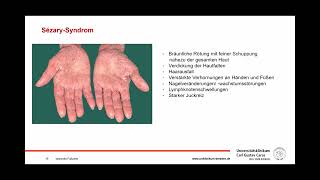
Other Signs. In addition to erythroderma and B2 blood involvement, patients with Sézary syndrome will typically have several other characteristic signs: generalized lymphadenopathy, opportunistic infections, and alopecia. The liver and possibly the spleen will be enlarged, and patients often have very thick, coarse skin on the soles of the feet and palms of the hands (i.e., palmoplantar keratoderma).
Diagnosis is usually made with a patient history, complete physical exam, blood tests, biopsy of skin lesions, computed tomography imaging, and sometimes lymph node biopsy and/or bone marrow biopsy. These methods can also be useful in determining the stage of disease, especially whether the lymph nodes have been involved and whether the cancerous cells have spread to blood and other organs. In addition to eczema and psoriasis, the differential diagnosis may include nonspecific dermatitis, lichen, lupus, pseudolymphoma, parapsoriasis, and toxidermia.


Информация по комментариям в разработке